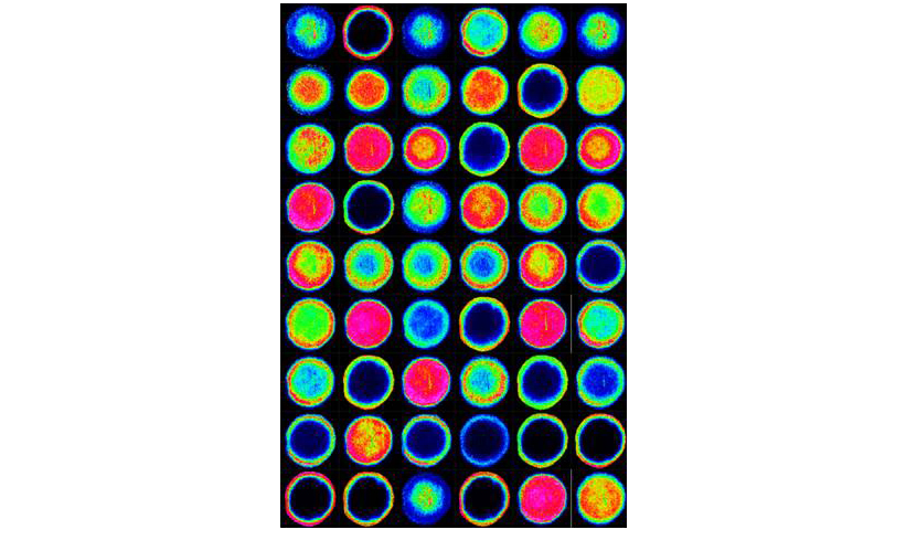
Shuman JHB, Lin AS, Truelock MD, Bryant KN, Piazuelo MB, Reyzer ML, Judd AM, McDonald WH, McClain MS, Schey KL, Algood HMS, Cover TL. Remodeling of the gastric environment in Helicobacter pylori-induced atrophic gastritis. mSystems, 9:e0109823, 2024.
Anderson DMG, Kotnala A, Migas LG, Patterson NH, Tideman L, Cao D, Adhikari B, Messinger JD, Ach T, Tortorella S, Van de Plas R, Curcio CA, Schey KL. Lysolipids are Prominent in Subretinal Drusenoid Deposits, a High-Risk Phenotype in Age-Related Macular Degeneration. Front. Ophthalmol., 3:1258734, 2023.
Wang Z, Gletten RB, Schey KL. Spatially Resolved Proteomics Reveals Lens Suture-Related Cell-Cell Junctional Protein Distributions. Invest. Ophthalmol. Vis Sci., 64:28, 2023.
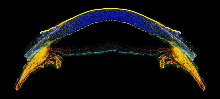
Lee HH, Tang Y, Bao S, Yang Q, Xu X, Schey KL, Spraggins JM, Huo Y, Landman BA. Unsupervised Registration Refinement for Generating Unbiased Eye Atlas. Proc SPIE Ent Soc Opt Eng, 12464:1246422, 2023.
Schey KL, Wang Z, Rose KL, Anderson DMG. Imaging Cataract-Specific Peptides in Human Lenses. Cells, 11:4042, 2022.
Wang Z, Cantrell LS, Schey KL. Spatially-Resolved Proteomic Analysis of the Lens Extracellular Diffusion Barrier. Invest. Ophthalmol. Vis. Sci., 62:25, 2021.
Cantrell LS and Schey KL. Data Independent Acquisition Mass Spectrometry of the Human Lens Enhances Spatiotemporal Measurement of Fiber Cell Aging. J Amer. Soc. Mass Spectrom., 32:2755-2765, 2021.
Giblin FJ, Anderson DMG, Han J, Rose KL, Wang Z, Schey KL. Acceleration of age-induced proteolysis in the guinea pig lens nucleus by in vivo exposure to hyperbaric oxygen: a mass spectrometry analysis. Exp. Eye Res., 210:108697, 2021.
Kotnala A, Anderson DMG, Patterson NH, Cantrell LS, Messinger JD, Curcio CA, Schey KL. Tissue fixation effects on human retinal lipid analysis by MALDI imaging and LC-MS/MS technologies. J Mass Spectrom., 56:e4798, 2021.
Guo G, Papanicolaou M, Demarais, NJ, Wang Z, Schey KL, Timpson P, Cox TR, Grey AC. HIT-MAP – a bioinformatics engine for automated annotation and visualisation of high-resolution spatial proteomic mass spectrometry imaging data. Nat. Commun., 12:3241, 2021.
Dufresne M, Fincher JA, Patterson NH, Schey KL, Norris JL, Caprioli RM, Spraggins JM. α-Cyano-4-hydroxycinnamic Acid and Tri-Potassium Citrate Salt Pre-Coated Silicon Nanopost Array Provides Enhanced Lipid Detection for High Spatial Resolution MALDI Imaging Mass Spectrometry. Anal. Chem., 93:12243-12249, 2021.
Lin A, Shuman J, Kotnala A, Shaw J, Beckett A, Harvey J, Tuck M, Dixon B, Reyzer M, Algood H, Schey K, Piazuelo M, Cover T. Loss of corpus-specific lipids in Helicobacter pylori-induced atrophic gastritis. mSphere, 6:0082621, 2021.
Grey AC and Schey KL. MALDI-MSI for Eye Diseases, in: MALDI MS Imaging: From Fundamentals to Spatial Omics, Porta T (Ed.), Royal Society of Chemistry, 2021, in press.
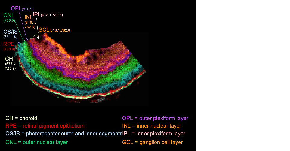
Anderson DMG, Messinger JD, Patterson NH, Rivera ES, Kotnala A, Spraggins JM, Caprioli RM, Curcio CA, Schey KL. Lipid landscape of the human retina and supporting tissues revealed by high-resolution imaging mass spectrometry. J. Amer. Soc. Mass Spectrom., 31:2426-2436, 2020.
Wang Z, Ryan DJ, Schey KL. Localization of the Lens Intermediate Filament Switch by Imaging Mass Spectrometry. Exp. Eye Res., 198:108134, 2020.
Anderson DM, Nye-Wood MG, Rose KL, Donaldson PJ, Grey AC, Schey KL. MALDI imaging mass spectrometry of b- and g-crystallins in the ocular lens. J. Mass Spectrom., 55:e4473, 2020.
Stark DT, Anderson DMG, Kwong JMK, Patterson NH, Schey KL, Caprioli RM, Caprioli J. Optic Nerve Regeneration after Crush Remodels the Injury Site: Molecular Insights from Imaging Mass Spectrometry. Invest Ophthalmol Vis Sci., 59:212-222, 2018.
Noble KV, Reyzer ML, Barth JL, McDonald H, Tuck M, Schey KL, Krug EL, Lang H. Use of Proteomic Imaging Coupled with Transcriptomic Analysis to Identify Biomolecules Responsive to Cochlear Injury. Front. Mol. Neurosci., 11:243, 2018.
Anderson DMG, Ablonczy Z, Koutalos Y, Hanneken AM, Spraggins JM, Calcutt W, Crouch RK, Caprioli RM, Schey KL. Bis(monoacyglycero)phosphate Lipids in the Retinal Pigment Epithelium Implicate Lysosomal/Endosomal Dysfunction in a Model of Stargardt Disease and Human Retinas. Sci. Reports 7:17352, 2017.
de Macedo CS, Anderson DM, Schey KL. MALDI (matrix assisted laser desorption ionization) Imaging Mass Spectrometry (IMS) of Skin: Aspects of Sample Preparation. Talanta 174:325-335, 2017.
Chen Y, Jester JV, Anderson DM, Marchitti SA, Schey KL, Thompson DC, Vasiliou V. Corneal Haze Phenotype in Aldh3a1-null Mice: In vivo Confocal Microscopy and Tissue imaging Mass Spectrometry. Chem. Biol. Interact. 276:9-147, 2017.
Anderson DMG, Calkins DJ, Ablonczy Z, Crouch RK, Caprioli RM, Schey KL. Imaging MS of Rodent Ocular Tissue and the Visual System, in: Imaging Mass Spectrometry – Methods and Protocols. Methods in Molecular Biology 1618:15-27, 2017.
Nye-Wood MG, Spraggins JM, Caprioli RM, Schey KL, Donaldson PJ, Grey AC. Spatial Distributions of Glutathione and Its Endogenous Conjugates in Normal Bovine Lens and a Model of Lens Aging. Exp. Eye Res. 154:70-78, 2016.
Gutierrez DB, Garland D, Schwacke JH, Hachey DL, Schey KL. Spatial Distributions of Phosphorylated Membrane Proteins Aquaporin 0 and MP20 Across Young and Aged Human Lenses. Exp. Eye. Res. 149:59-65, 2016.
Schey KL, Hachey AJ, Rose KL, Grey AC. MALDI Imaging Mass Spectrometry of Pacific White Shrimp L. vannamei and Identification of Abdominal Muscle Proteins. Proteomics, 16:1767-1774, 2016.
Wenke JL, McDonald WH, Schey KL. Spatially-Directed Proteomics of the Human Lens Outer Cortex Reveals an Intermediate Filament Switch Associated with the Remodeling Zone. Invest. Ophthalmol. Vis. Sci., 57:4108-4114, 2016.
Bowery HE, Anderson DM, Pallitto P, Gutierrez DB, Fan J, Crouch RK, Schey KL, Ablonczy Z. Imaging mass spectrometry of the visual system: advancing the molecular understanding of retina degenerations. Proteom Clin Appl., 10:391-402, 2016.
Anderson DMG, Van de Plas R, Rose KL, Hill S, Schey KL, Solga AC, Gutmann DH, Caprioli RM. 3-D Imaging Mass Spectrometry of Protein Distributions in Mouse Neurofibromatosis 1 (NF1)-Associated Optic Glioma. J. Proteomics, S1874-3919:30027-6, 2016.
Anderson DMG, Floyd KA, Barnes S, Clark JM, Clark JI, Mchaourab H, Schey KL. A method to prevent protein delocalization in imaging mass spectrometry of non-adherent tissues: application to small vertebrate lens imaging. Anal. Bioanal. Chem. 407:2311-2320, 2015.
Anderson DMG, Spraggins J, Rose KL, Schey KL. High Spatial Resolution Imaging Mass Spectrometry of Human Optic Nerve Lipids and Proteins. J. Amer. Soc. Mass Spectrom. 26:940-947, 2015.
de Macedo CS, Anderson DM, Pascarelli BM, Spraggins JM, Sarno EN, Schey KL, Pessolani MCV. MALDI Imaging Reveals Lipid Changes in the Skin of Leprosy Patients Before and After Multidrug Therapy (MDR). J. Mass Spectrom. 50:1374-1385, 2015.
Adler L, Boyer NP, Anderson DM, Spraggins JM, Schey KL, Hanneken A, Ablonczy Z, Crouch RK, Koutalos Y. Determination of N-retinylidene-N-retinylethanolamine (A2E) Levels in Central and Peripheral Areas of Human Retinal Pigment Epithelium. Photochem. Photobiol. Sci., 14:1983-1990, 2015.
Patel S, Ling J, Kim SJ, Schey KL, Rose K, Kuchtey RW. Proteomic Analysis of Macular Fluid Associated with Advanced Glaucomatous Excavation. JAMA Opthalmol., 5:1-3, 2015.
Wenke JL, Rose KL, Spraggins JM, Schey KL. MALDI Imaging Mass Spectrometry Spatially Maps Age-Related Deamidation and Truncation of Human Lens Aquaporin-0. Invest. Ophthalmol. Vis. Sci., 56:7398-7405, 2015.
Anderson DMG, Ablonczy A, Koutalos Y, Spraggins J, Crouch RK, Caprioli RM, Schey KL. High Resolution MALDI- Imaging Mass Spectrometry of Retinal Tissue Lipids. J. Amer. Soc. Mass Spectrom. 25:1394-1403, 2014.
Ablonczy Z, Smith N, Anderson DM, Grey AC, Spraggins J, Koutalos Y, Schey KL, Crouch RK. The utilization of fluorescence to identify the components of lipofuscin by imaging mass spectrometry. Proteomics 14:936-944, 2014.
Nicklay JJ, Harris GA, Schey KL, Caprioli RM, MALDI imaging and in situ identification of integral membrane proteins from rat brain tissue sections, Anal Chem, 85:7191-7196, 2013.
Ablonczy Z, Higbee D, Grey AC, Koutalos Y, Schey KL, Crouch RK. Similar molecules spatially correlate with lipofuscin and A2E in the mouse but not in the human RPE. Arch. Biochem. Biophys., 539:196-202, 2013.
Ablonczy Z, Higbee D, Anderson DM, Dahrouj M, Grey AC, Koutalos Y, Schey KL, Gutierrez D, Hanneken A, Crouch RK. Lack of Correlation between the Spatial Distribution of A2E and Lipofuscin Fluorescence in the Human Retinal Pigment Epithelium. Invest. Ophthalmol. Vis. Sci, 54:5535-542, 2013.
Anderson DMG, Mills D, Spraggins J, Lambert WS, Calkins DJ, Schey KL. High Resolution MALDI-Imaging Mass Spectrometry of Lipids in Rodent Optic Nerve Tissue, Mol. Vis. 19:581-592, 2013.
Schey KL, Anderson DMG, Rose KL. Spatially-Directed Protein Identification from Tissue Sections by Top-Down LC-MS/MS with Electron Transfer Dissociation. Anal. Chem., 85:6767-6774, 2013.
Wang Z, Han J, David LL, Schey KL. Proteomics and Phosphoproteomics Analysis of Human Lens Fiber Cell Membranes, Invest. Ophthalmol. Vis. Sci. 54:1135-1143, 2013.
Schey KL, Grey AC, Nicklay JJ. Mass Spectrometry Analysis of Membrane Proteins: A Focus on Aquaporins, Biochemistry, 52:3807-3817, 2013.
Grey, AC, Walker KL, Petrova RS, Han J, Wilmarth PA, David LL, Donaldson PJ, Schey KL. Verification and Spatial Localization of Aquaporin-5 in the Ocular Lens. Exp. Eye Res. 108:94-102, 2013.
Ablonczy Z, Gutierrez DB, Grey AC, Schey KL, Crouch RK. Molecule-specific imaging and quantitation of A2E in the RPE. Adv. Exp. Med. Biol. 723:75-71, 2012.
Gutierrez DB, Garland D, Schey KL. Spatial analysis of human lens aquaporin-0 post-translational modifications by MALDI mass spectrometry tissue profiling, Exp. Eye Res., 93:912-920, 2011.
Qureshi A, Grey A, Rose KL, Schey KL, Del Poeta M. Cryptococcus neoformans modulates extracellular killing by neutrophils. Frontiers in Microbiol. 2:193, 2011.
Grey AC, Crouch RK, Koutalos Y, Schey KL, and Ablonczy Z. Spatial Localization of A2E in the Retinal Pigment Epithelium, Invest. Ophthalmol. Vis. Sci. 52:3926-3933, 2011.
Qureshi A, Subathra M, Grey A, Schey K, Del Poeta M, Luberto C. Role of sphinghomyelin synthase in controlling the antimicrobial activity of neutrophils against Cryptococcus neoformans. PLoS One, 5:e15587, 2010.
Stella DR, Floyd KA, Grey AC, Renfrow MB, Schey KL, Barnes S. Tissue localization and solubilities of αA-crystallin and its numerous C-terminal truncation products in pre- and post-cataractous ICR/f rat lenses. Invest. Ophthalmol. Vis. Sci., 51:5153-5161, 2010.
Uys JD, Grey AC, Wiggins A, Schwacke JH, Schey KL, Kalivas PW. Matrix-Assisted Laser Desorption/Ionization Tissue Profiling of Secretoneurin in the Nucleus Accumbens Shell from Cocaine Sensitized Rats. J. Mass Spectrom., 45:97-103, 2010.
Grey AC, Gelasco AK, Moreno-Rodriguez RA, Krug EL, Schey KL. Molecular Morphology of the Chick Heart Visualized by MALDI Imaging Mass Spectrometry. Anat. Rec., 293:821-8, 2010.
Grey AC and Schey KL. Age-related Changes in the Spatial Distribution of Human Lens α-Crystallin Products by MALDI Imaging Mass Spectrometry, Invest. Ophthalmol. Vis. Sci., 50:4319-4329, 2009.
Grey AC, Chaurand P, Caprioli, RM , Schey KL. MALDI Imaging Mass Spectrometry of Integral Membrane Proteins from Ocular Lens and Retinal Tissue, J. Proteom. Res., 8:3278-3283, 2009.
Grey AC and Schey KL. Distribution of Bovine and Rabbit Lens α-Crystallin Products by MALDI Imaging Mass Spectrometry, Mol. Vis., 2008, 14:171-9.
Thibault DB, Gillam CJ, Han J, Schey KL. MALDI Tissue Profiling of Integral Membrane Proteins from Ocular Tissues, J. Amer. Soc. Mass Spectrom., 19:814-22, 2008.
Wang Z, Han J, Schey KL. Spatial Differences in an Integral Membrane Proteome Detected in Laser Capture Microdissected Samples, J. Proteome Res., 7:2696-702, 2008.
Han J and Schey KL. MALDI Tissue Imaging of Ocular Lens α-Crystallin, Invest Ophthalmol Vis Sci, 2006, 47:2990-6.


